About x-rays
DescriptionAn x-ray creates a picture of the inside of the body by using a small amount of radiation. This radiation is absorbed differently by various parts of the body, such as bones and soft tissues, allowing them to be visualised non-invasively. X-rays can be used to diagnose broken bones, identify foreign objects, and examine other potential injuries or infections. Example of UsesX-ray exams can be used to view, monitor, or diagnose the following:
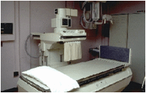
Preparation for an x-ray examYou will be asked to remove metal objects before the test. If your clothing has metal on it, like a zip, you will be asked to change into a patient gown. During the ExamYou will be asked to either lie down on an exam table or stand. The room may be cool in order to keep the equipment from overheating. You may be asked to hold very still, without breathing for a few seconds. The technologist will step behind a radiation barrier and activate the x-ray machine. Often multiple images or views are taken from different angles, so the technologist will reposition you for another view and the process will be repeated. You will not feel the radiation.
BenefitsX-ray exams are non-invasive, quick, and easy. The equipment used is relatively inexpensive and widely available. RisksX-ray exams exposure patients to radiation. The amount of radiation exposure is variable depending upon the x-ray type (for example, lungs or bones) and the x-ray machine type (for example, different models and manufacturers). Because the radiation exposure is variable, the risks are also variable. Please speak to your referring practitioner for specific details on radiation exposure and possible risks. Women need to inform their doctor if they are or may be pregnant prior to any radiological imaging. Your doctor may recommend another type of test to reduce the possible risk. ResultsA radiologist will analyse and interpret the results of your x-ray. The report will be sent to your referring practitioner the following day. The radiology report is meant to be read in conjunction with the clinical assessment of the referring clinician. Please obtain your imaging report from your referring practitioner.
|
||||||
Bibliography
http://www.acrin.org/PATIENTS/ABOUTIMAGINGEXAMSANDAGENTS/ABOUTXRAYS.aspx

